What Are Lymphedema Wounds Called?

In medical language, wounds caused by long-standing lymphedema are most often called lymphostatic ulcers or lymphedematous ulcers. In people who also have vein disease (which is very common), those open wounds are usually classified as venous leg ulcers that occur in the setting of lymphedema or phlebolymphedema (combined venous + lymphatic disease).
1. Quick refresher: what is lymphedema?
Lymphedema is chronic swelling caused by failure of the lymphatic system, leading to protein-rich fluid, low-grade inflammation, and progressive fibrosis in the tissues.
Over time, especially in the legs, this constant swelling can:
- Stretch and thin the skin
- Reduce oxygen delivery and waste removal
- Increase the risk of infection (cellulitis)
- Make even small injuries very slow to heal
That’s why lymphedema and leg ulcers often show up together.
2. What are lymphedema wounds actually called?
2.1 Lymphostatic ulcers / lymphedematous ulcers
Chronic ulcers that form because of lymphatic failure are commonly referred to as:
- Lymphostatic ulcers
- Lymphedematous ulceration
These terms highlight that the primary problem is lymphatic stasis — lymph fluid stagnating in the tissues, damaging skin and micro-circulation.
Typical features described in the literature include:
- Long-standing leg swelling, often with “woody” fibrosis
- Thickened, scaly or warty skin changes (hyperkeratosis, papillomatosis)
- Lymphorrhea – clear lymph fluid leaking through tiny breaks (“weeping legs”)
- Shallow, irregular ulcers with lots of exudate and macerated surrounding skin
- Recurrent infections (cellulitis, erysipelas)
Only a subset of people with lymphedema will develop these ulcers, but when they do, they are typically hard to heal and easy to re-open.
2.2 Venous leg ulcers in phlebolymphedema
In real-world practice, many “lymphedema wounds” are actually mixed venous + lymphatic ulcers:
- Chronic venous insufficiency damages the veins.
- The overwhelmed lymphatic system can’t clear the extra fluid → phlebolymphedema.
Open wounds in this scenario are almost always documented as venous leg ulcers (VLUs), even though lymphatic failure is heavily involved.
So a patient might hear several phrases for “the same” wound:
- Venous leg ulcer with lymphedema
- Venous stasis ulcer in a lymphedematous leg
- Phlebolymphedema with VLU
The label matters for coding and guidelines, but for patients the key point is that both the veins and lymph system need attention, not just the open sore.
2.3 Other wound types that appear on lymphedema limbs
A major taxonomy paper on lymphedema skin disorders lists a range of wounds that can occur on lymphedema-affected limbs:
- Non-healing surgical wounds
- Traumatic wounds (cuts, falls, minor injuries that never quite close)
- Malignant or irradiated wounds
- Venous stasis ulcers / phlebolymphedema ulcers
In these cases, the wound is named by the original cause (surgical, malignant, traumatic), but lymphedema is a major reason it heals so poorly.
3. How lymphedema turns into chronic ulcers
Several mechanisms link lymphedema to hard-to-heal wounds:
-
Protein-rich edema
Lymph fluid is full of protein. When it sits in the tissues, it fuels chronic inflammation and fibrosis, damaging the micro-circulation. -
Skin barrier breakdown
Stretching, dryness, and thickening (hyperkeratosis) make the skin fragile. Small scratches or fungal infections between toes can evolve into big ulcers. -
Lymphorrhea (“weeping skin”)
Clear lymph fluid leaks through tiny cracks or blisters. Constant moisture macerates the skin, making it even easier for an ulcer to appear. -
Infection cycle
Bacteria love warm, moist, protein-rich environments. Recurrent cellulitis further damages lymphatics, creating a vicious circle. -
Reduced oxygen delivery
Fibrosis and edema physically increase the distance between blood vessels and skin cells, starving tissues of oxygen and nutrients.
When a break in the skin happens in this environment, we get a chronic lymphedema ulcer rather than a simple cut that heals in a week.
4. What do lymphostatic / lymphedema ulcers look like?
Descriptions from wound and lymphedema resources highlight several common patterns, especially on the lower legs and ankles:
- Location: often around the gaiter area (above the ankle), on swollen, fibrotic legs
- Shape: shallow, irregular edges, sometimes multiple small ulcers that merge
- Exudate: heavy drainage of lymph-rich fluid; surrounding skin is soggy and white (macerated)
- Skin: thick, bumpy, discolored (brown, red, or “rusty”), with scaling or papillomatosis
- Symptoms: heaviness, aching, itching; high risk of recurrent infection
Because these ulcers sit on a limb that is already swollen and heavy, they can be very disabling—people struggle with walking, work, shoes, and sleep.
5. Why the exact name matters (and when it doesn’t)
For clinicians and insurers, calling a wound a venous leg ulcer, lymphostatic ulcer, or traumatic non-healing wound points to:
- Different guideline pathways
- Different compression protocols
- Different expectations about healing time and recurrence
For patients, the key is simpler:
- “I have chronic lymphedema.”
- “I have an open leg ulcer/wound on top of that.”
- “Both the swelling and the wound need to be treated together.”
Whether your report says “venous ulcer,” “lymphostatic ulcer,” or “phlebolymphedema with VLU,” you’re dealing with a high-risk chronic wound in an edematous limb that deserves specialist attention.
6. How lymphedema-related ulcers are managed (in broad strokes)
Guidelines and reviews on lymphedema wound care all stress a combined approach: treat the wound and the swelling.
6.1 Control the swelling
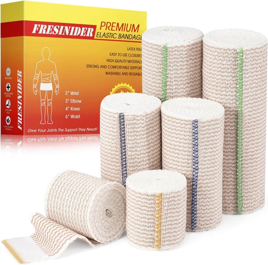
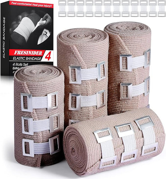
- Compression therapy (elastic bandages, multilayer wraps, or compression garments)
- Complete Decongestive Therapy (CDT) – manual lymph drainage, compression, exercise, and skin care
- Elevation and movement to support venous and lymphatic return
6.2 Protect the skin and wound
- Use absorbent, non-adherent dressings (e.g., foam dressings, super-absorbents) to manage heavy lymphorrhea and prevent maceration.
- Treat fungal infections and eczema around the wound to protect the barrier.
- Moisturize intact skin daily to reduce cracking.
6.3 Prevent and treat infection
- Watch closely for cellulitis (sudden redness, heat, pain, fever).
- Use topical or systemic antibiotics when indicated, as directed by a clinician.
6.4 Education and long-term maintenance
Patients are often taught how to change dressings, wrap legs, inspect skin, and know when to call for help. Because the underlying lymphedema doesn’t “go away,” ongoing compression and skin care are essential even after an ulcer has healed.
7. When to seek urgent medical help
Anyone with lymphedema should contact a healthcare professional promptly if they notice:
- A new open area, blister, or ulcer that doesn’t improve within a few days
- Sudden increased redness, heat, or pain in the limb
- Fever, chills, or feeling very unwell
- Rapidly spreading rash or red streaks (possible cellulitis)
Early treatment can prevent a small lymphostatic ulcer from turning into a large, chronic wound that dominates daily life.
Key takeaways
- Wounds that develop in chronically swollen, lymphedematous limbs are commonly called lymphostatic ulcers or lymphedematous ulcers.
- When venous disease is also present (phlebolymphedema), they’re usually labelled venous leg ulcers in a lymphedema limb.
- These ulcers are driven by protein-rich edema, skin changes, lymphorrhea, and recurrent infection, making them hard to heal.
- Effective care has to address both the open wound and the underlying swelling with compression, skin care, and infection control, ideally under a specialist team.
For readers with lymphedema, the most important step isn’t memorizing the exact term — it’s recognizing skin changes early and getting timely, expert help so those “lymphedema wounds” have the best chance to heal.